Learn vocabulary terms and more with flashcards games and other study tools. Reset Help Fibula Lateral meniscus Anterior cruciate ligament Ligaments That Stabilize the Knee Joint Tbial collateral ligament Articular cartilage Fibular collateral ligament Patellar surface Tibia Patelar igament cut Posterior cruciate ligament Medial meniscus.

Solved Art Labeling Activity Structural Features Chegg Com
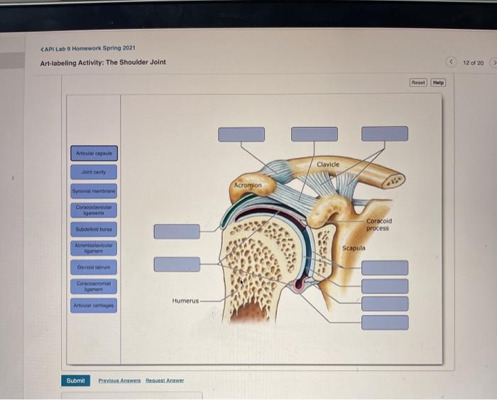
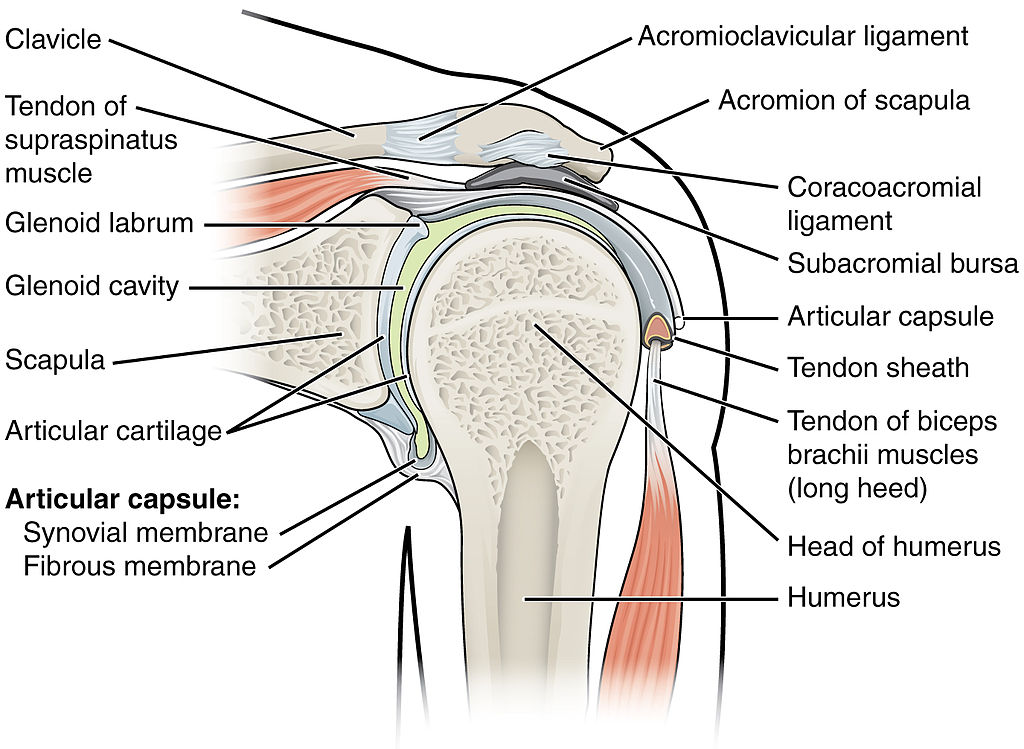
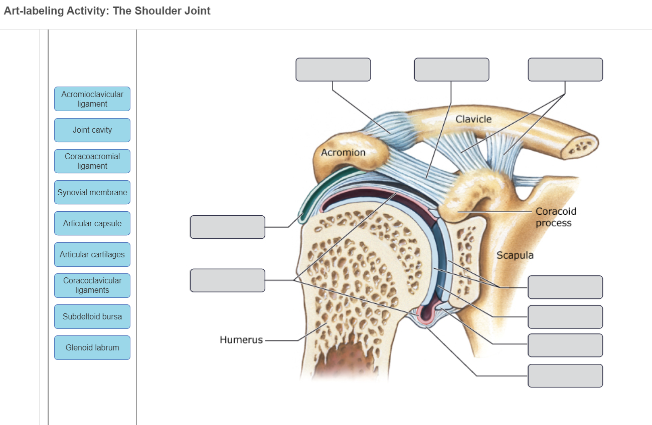
The Shoulder Joint Acromioclavicular ligament Clavicle Joint cavity Acromion Coracoacromial ligament Synovial membrane Coracoid process Articular capsule Articular cartilages Scapula Coracoclavicular ligaments Subdeltoid bursa Humerus Glenoid labrum.
. Figure 233a 1 of 2 Art-labeling Activity. The right elbow joint lateral view Sets found in the same folder. To learn the curves and regions of the vertebral column.
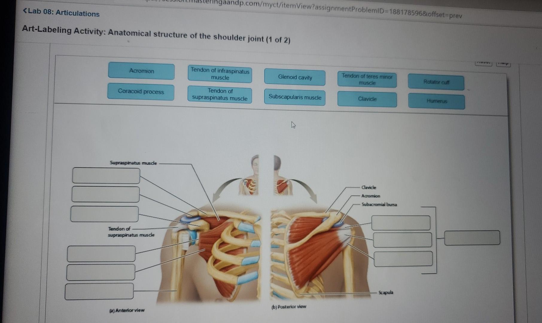
Structural features ligaments and associated tendons of the shoulder joint. Word bank is below image. Structural features ligaments and associated tendons of the shoulder joint Drag the labels onto the diagram to identify the struclural features ligaments_ and associaled tendons of the shoulder join Acomioclavicular Ilgament Glenohumeral ligaments Glenoid cavily Glenoid labrum Tendon 0f biceps brachil muscle Aricular capsule Coracohumera Iigament.
Anatomy and Physiology questions and answers. The Structure of a Synovial Joint. Structural features ligaments and associated tendons of the shoulder joint Glenoid cavity Articular capsule Glenoid labrum 1 Glenohumeral ligaments Tendon of biceps brachii muscle W Acromioclavicular ligament Coracohumeral ligament Submit Previous Answer.
Structural features ligaments and associated tendons of the shoulder joint. Art-labeling Activities Use the art-labeling activities to quiz yourself on key anatomical structures in this chapter. Shoulder and Back Deep Dissectionjpg.
Angular Movements of the Joints. Chapter Test - Chapter 8 Question 3. Miami Dade College.
M no subject - stellarvore91gm x G anatomy abdominal quadrants - X Course Home X. Each year the American Heart Association AHA in conjunction with the Centers for Disease Control and Prevention the National Institutes of Health and other government agencies brings together. Learn vocabulary terms and more with flashcards games and other study tools.
Muscles of the upper limb. Muscles that move the hand and fingers posterior view middle layer. Pictures of joint movements.
Part A Drag the labels onto the diagram to identify the. Structure of a Synovial Joint Label the structures of a synovial joint. This muscular system picture shows all the major muscle groups on the human body from the frontal view.
Start studying Art-labeling Activity. Pictures of joint movements. The right elbow joint medial view.
The Right Elbow Joint Showing Stabilizing Ligaments. Apr 06 2022 0802 AM. Grading Policy Art-labeling Activity.
Start studying Art-labeling Activity. Anatomy and Physiology. Lecture Exam 1- AP Semester 1.
To see a muscular system diagram from the posterior back view click here. Part A Drag the correct label to the appropriate location to identify the structures of a synovial joint. Anatomy and Physiology questions and answers.
Chapter 73 Anatomy module. 1059pm on Sunday February 7 2021 You will receive no credit for items you complete after the assignment is due. Help labeling the bones and synovial joints of the body by looking at the image and providing the bone AND synovial joint type for each number.
Muscles of the Neck Shoulder and Back Deep Dissectionjpg. Structural features ligaments and associated tendons of the shoulder joint. A shoulder separation that involves the lateral end of the clavicle sliding onto the superior aspect of the acromion would.
View art labeling activity - vertebral anatomyjpg from BSC MISC at Miami Dade College Miami. Jones PE Water Agency Director 660 South Cobb Drive Marietta GA 30060 770 419-6200 770 419-6224 Fax Find us on Nextdoor. To learn the angular movements of the joints.
The Knee Joint Drag the correct label to the appropriate structure of the knee joint. The right elbow joint lateral view PICTURE. Rotational Movements of the Joints.
View art labeling activity -structure of a nail superficial and cross-sectional viewsjpg from BSC MISC at Miami Dade College Miami. Week 2 Chapter 8_ Due. Curves and Regions of the Vertebral Column Learning Goal.
View art labeling activity - anatomy of a thoracic vertebrajpg from EGN MISC at Miami Dade College Miami. The Shoulder Joint Acromioclavicular Ligament Clavicle Joint Cavity Acromion Coracoacromial Ligament. 1159pm on Friday October 6 2017 To understand how points are awarded read the Grading Policy for this assignment.
The right elbow joint medial view. Synovial joint components sagittal section PICTURE. Start studying the Art- labeling Activity flashcards containing study terms like and more.
Drag the appropriate labels to their respective targets. Label the curves and regions of the vertebral column. The shoulder girdle is also called the pectoral girdle and it is a bone ring incomplete posteriorlyThe shoulder girdle is formed by two sets of bones.
Structural features ligaments and associated tendons of the shoulder joint Art-labeling Activity. Identify and test your knowledge of the locations of the muscles of the torso with this great free quiz - Anterior locations Quiz 2.
Solved Chegg Com
File 914 Shoulder Joint Jpg Wikimedia Commons

Art Labelling Study Set Flashcards Quizlet

Shoulder Joint Label Pictures Flashcards Quizlet
Solved Art Labeling Activity The Shoulder Joint Chegg Com
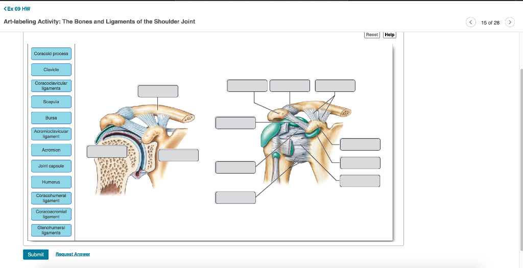
Solved Ex 09 Hw Art Labelling Activity The Bones And Chegg Com
Solved Capi Lab 9 Homework Spring 2021 Art Labeling Chegg Com
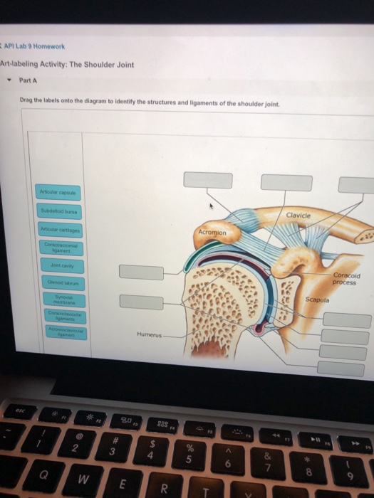
Api Lab 9 Homework Art Labeling Activity The Chegg Com
0 comments
Post a Comment